THE SCIENTIFIC DISCOVERY JOURNAL · OPTICS & MICROSCOPY
The Invention That Revealed an Invisible World
From simple magnifying lenses to the discovery of cells and microorganisms, the microscope transformed how scientists understand living things.
For thousands of years, people studied the natural world using only what their eyes could see. They observed plants, animals, human anatomy, and the movement of celestial objects, but an enormous part of nature remained hidden.
Individual cells, bacteria, and many microscopic structures were too small to observe directly.
The development of the microscope changed that limitation.
By using carefully shaped lenses to magnify tiny objects, early investigators began discovering structures that had never been described before.
What initially appeared to be a useful optical instrument gradually became one of the most important tools in scientific history.
The microscope transformed biology by revealing that living organisms contain microscopic structures and that an extraordinary variety of life exists beyond ordinary human vision.
Its development helped establish cell biology, microbiology, and new approaches to medical research.
Life Before the Microscope
Before microscopes became available, natural philosophers and physicians could examine only structures visible to the unaided eye.
They could study organs, bones, leaves, and other relatively large features.
However, the detailed organization of tissues remained difficult to understand.
People knew that diseases could spread, but they lacked the tools needed to observe bacteria and many other microorganisms.
The internal organization of plants and animals was also largely unknown.
Although magnifying lenses had been used for centuries, they did not immediately lead to systematic microscopic biology.
Scientists needed instruments capable of producing useful magnification and sufficient image clarity.
Advances in lens-making eventually made that possible.
How Early Lenses Led to the Microscope
The history of microscopy begins with the development of optical lenses.
A convex lens bends incoming light in ways that can make nearby objects appear larger.
Magnifying glasses demonstrated this principle long before the invention of the compound microscope.
During the late sixteenth and early seventeenth centuries, European instrument makers experimented with combining lenses inside tubes.
These experiments contributed to the development of early compound microscopes.
The precise identity of the first inventor remains debated.
Members of the Janssen family are often associated with early microscope development, but surviving historical evidence does not establish a single uncontested inventor.
Early microscopes also suffered from optical limitations.
Images could appear distorted, poorly illuminated, or surrounded by unwanted colors.
Nevertheless, the instruments demonstrated that magnification could reveal details invisible to the naked eye.
Simple and Compound Microscopes: What’s the Difference?
Early microscopy developed along two important approaches.
A simple microscope uses a single lens or lens system to magnify an object.
A compound microscope uses multiple lens systems, typically including an objective lens and an eyepiece.
The objective produces an enlarged image, while the eyepiece allows the observer to view it at additional magnification.
However, magnification alone does not determine image quality.
Resolution—the ability to distinguish closely spaced details—is equally important.
A microscope that enlarges a blurry image does not necessarily reveal additional useful information.
Early researchers therefore faced challenges involving lens quality, lighting, and optical distortion.
OPTICAL INSTRUMENTS / COMPARATIVE NOTES
Uses a single magnifying lens or optical assembly. Carefully made simple microscopes can produce highly useful images.
Uses multiple lens systems to magnify specimens and became central to modern laboratory microscopy.
Robert Hooke and the Discovery of Cells
One of the most influential early microscope users was English scientist Robert Hooke.
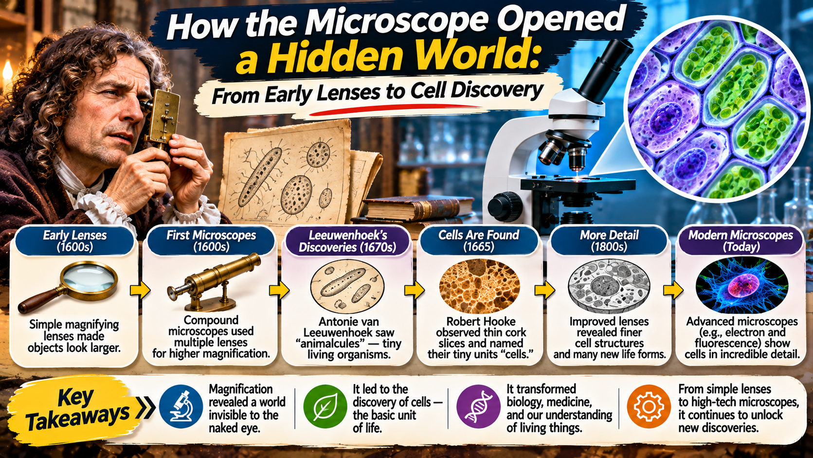
In 1665, Hooke published Micrographia, a remarkable illustrated work describing observations made with magnifying instruments.
The book included detailed images of insects, plant materials, and other small structures.
Among Hooke’s most famous observations was a thin section of cork.
Under magnification, the cork appeared to contain many small compartments.
Hooke called these structures “cells” because they reminded him of small rooms.
The structures he observed were primarily the walls of dead plant cells.
He did not discover the modern biological concept of the cell in its complete form.
However, his terminology became foundational to later biological research.
Micrographia also helped demonstrate the scientific value of detailed microscopic observation.
HISTORICAL LABORATORY NOTE · 1665
Robert Hooke Named the Tiny Compartments He Observed in Cork “Cells.”
The name endured, even though scientists would need nearly two more centuries to develop a comprehensive theory of cells as the fundamental units of living organisms.
Antonie van Leeuwenhoek and the Discovery of Microorganisms
Another major figure in early microscopy was Dutch investigator Antonie van Leeuwenhoek.
He developed exceptional skill in making and using small, powerful single-lens microscopes.
His instruments allowed him to observe structures that many contemporary microscopes could not resolve clearly.
During the 1670s, Leeuwenhoek described microscopic organisms living in water and other materials.
He referred to some of these tiny living forms as “animalcules.”
His observations included microorganisms that would later be recognized as belonging to groups such as protozoa and bacteria.
Leeuwenhoek communicated many of his discoveries through letters to the Royal Society in London.
These reports expanded scientific awareness of microscopic life.
His work demonstrated that water and other seemingly ordinary materials could contain living organisms invisible to the naked eye.
This discovery fundamentally changed ideas about the diversity of life.
A Timeline of Important Microscope Discoveries
SCIENTIFIC DISCOVERY TIMELINE
Early compound microscope designs emerge through experiments with multiple lenses.
Robert Hooke publishes Micrographia and introduces the term “cells” for structures observed in cork.
Antonie van Leeuwenhoek reports observations of microscopic living organisms.
Matthias Schleiden and Theodor Schwann help establish cell theory.
Electron microscopy and advanced optical methods reveal increasingly detailed cellular structures.
How Microscopes Helped Establish Cell Theory
Hooke’s observations introduced the term “cell,” but scientists did not immediately understand the biological importance of these structures.
During the nineteenth century, improved microscopes allowed researchers to examine plant and animal tissues in greater detail.
In 1838, Matthias Schleiden argued that plants were composed of cells.
In 1839, Theodor Schwann extended important cellular principles to animals.
Their work helped establish the foundations of cell theory.
Later research strengthened the understanding that new cells arise from existing cells.
Cell theory became one of the central principles of modern biology.
It explains that living organisms are composed of cells and that cells are fundamental units of biological organization.
Microscopy was essential to developing this understanding because cells could not be studied adequately through ordinary observation alone.
Why Better Lenses Made Better Discoveries Possible
Early microscopes often produced images affected by optical distortion.
Chromatic aberration occurs when different wavelengths of light focus at different positions, creating unwanted color effects.
Spherical aberration can also reduce image sharpness.
During the eighteenth and nineteenth centuries, advances in lens manufacturing helped reduce these problems.
Improved objective lenses, illumination systems, and mechanical designs made microscopes more reliable scientific instruments.
Better resolution allowed researchers to distinguish finer details in biological specimens.
These improvements supported the growth of histology, the microscopic study of tissues, and other specialized fields.
Microscopes gradually became essential equipment in laboratories, universities, and medical institutions.
How Microscopy Changed Medicine
The microscope transformed medicine by allowing researchers to investigate structures and organisms that could not be seen directly.
Scientists could examine blood cells, tissue organization, and microscopic changes associated with disease.
Microscopy also supported the development of microbiology.
During the nineteenth century, researchers including Louis Pasteur and Robert Koch helped establish important connections between microorganisms and infectious diseases.
The microscope was one of several essential tools used in this work.
It allowed investigators to observe microorganisms and examine biological samples.
Combined with laboratory culture methods, experimental research, and improved staining techniques, microscopy contributed to major advances in understanding infection.
Modern medical laboratories continue to use microscopes for examining cells, tissues, microorganisms, and other diagnostic materials.
From Light Microscopes to Electron Microscopes
Traditional light microscopes use visible light and optical lenses to produce magnified images.
They remain valuable for observing many biological structures.
However, the wavelength of visible light limits the smallest details that conventional optical microscopes can resolve.
During the twentieth century, electron microscopes introduced a different approach.
Instead of visible light, they use beams of electrons and electromagnetic lenses.
Because electrons can have much shorter effective wavelengths than visible light, electron microscopes can reveal extremely fine structures.
Transmission electron microscopes are especially useful for studying thin internal structures.
Scanning electron microscopes can produce detailed information about specimen surfaces.
These instruments have transformed research into cells, materials, and microorganisms.
However, electron microscopy often requires specialized specimen preparation and operating conditions.
Comparing Major Types of Microscopes
| Microscope Type | Main Technology | Typical Applications |
|---|---|---|
| Simple microscope | Single magnifying lens system | Magnifying small objects and details |
| Compound light microscope | Visible light and multiple lenses | Cells, tissues, and microorganisms |
| Fluorescence microscope | Excitation and detection of fluorescent light | Labeled molecules and cellular structures |
| Transmission electron microscope | Electron beam passing through thin specimens | Fine internal structures |
| Scanning electron microscope | Focused electron beam scanning a surface | Detailed surface structures |
What Scientists Can See with Modern Microscopes
Modern microscopy allows researchers to investigate biological structures at many different scales.
Light microscopes can reveal cell shapes, tissue organization, and the movement of certain microorganisms.
Fluorescence microscopy allows scientists to highlight selected molecules or cellular components using fluorescent markers.
Advanced optical techniques can produce detailed images of living cells and biological processes.
Electron microscopy provides access to much finer structural information.
Researchers use these methods to study cellular membranes, organelles, microorganisms, and the organization of biological materials.
Different instruments are chosen according to the scientific question.
No single microscope is ideal for every type of specimen or investigation.
Why the Microscope Remains Essential Today
The microscope continues to support research across numerous scientific disciplines.
Biologists use microscopy to study cells and tissues.
Medical professionals examine biological samples to investigate disease.
Environmental scientists observe microorganisms in water, soil, and other ecosystems.
Materials scientists study microscopic structures that influence the properties of metals, polymers, and other substances.
Modern microscopy also benefits from digital imaging and computational analysis.
Researchers can capture detailed images, measure structures, and compare observations using specialized software.
These developments have expanded the microscope’s usefulness far beyond the capabilities of early optical instruments.
Nevertheless, the fundamental scientific purpose remains familiar: making structures accessible to observation that would otherwise remain hidden.
Frequently Asked Questions
Who invented the microscope?
The microscope developed through contributions from multiple instrument makers and scientists. The identity of the first inventor remains historically debated.
Who discovered cells?
Robert Hooke introduced the term “cells” in 1665 after observing the compartment-like structure of cork under a microscope.
Who first observed microorganisms?
Antonie van Leeuwenhoek made pioneering observations of microscopic living organisms during the seventeenth century.
What is the difference between magnification and resolution?
Magnification makes an image appear larger, while resolution determines how clearly closely spaced details can be distinguished.
How did microscopes help develop cell theory?
Improved microscopes allowed scientists to examine plant and animal tissues and recognize cells as fundamental components of living organisms.
Why are electron microscopes more powerful than ordinary light microscopes?
Electron microscopes use electron beams that can provide much finer structural resolution than conventional visible-light microscopy.
Are microscopes still important in modern medicine?
Yes. Microscopy remains important for examining cells, tissues, microorganisms, and many diagnostic specimens.
Final Thoughts
The invention and development of the microscope fundamentally changed humanity’s understanding of the natural world.
Early lenses introduced the possibility of magnifying tiny objects, while improved instruments allowed investigators to observe structures that had never been described before.
Robert Hooke’s observations helped establish the terminology of cells, and Antonie van Leeuwenhoek revealed the existence of microscopic living organisms.
Later advances in optical technology supported the development of cell theory, microbiology, and modern medical research.
Electron microscopy and advanced imaging techniques have since expanded scientific observation to increasingly fine scales.
The microscope remains one of the clearest examples of how a technological invention can transform scientific knowledge.
By revealing a hidden world of cells and microorganisms, it changed not only what scientists could see, but also how they understood life itself.